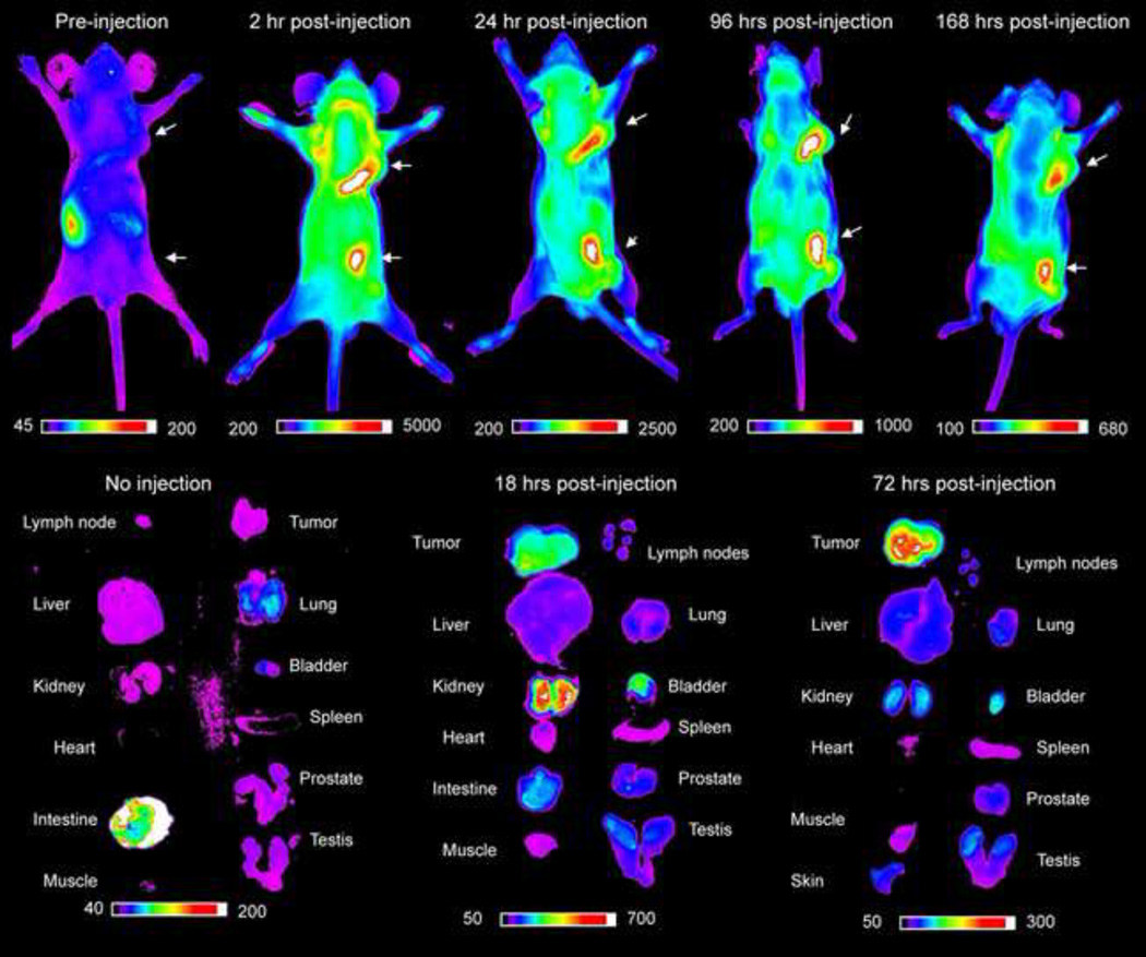
Figure 3.
In vivo imaging of canine melanoma xenograft tumors in nude mice with OA02-Cy5.5. The color scales represent fluorescent intensity detected with optical imaging, with the greatest fluorescence to the right of the scale. There is a high uptake of peptide in the tumor tissue in whole body images and ex-vivo images. The tumor remains strongly fluorescent up to 168 hours after injection.

