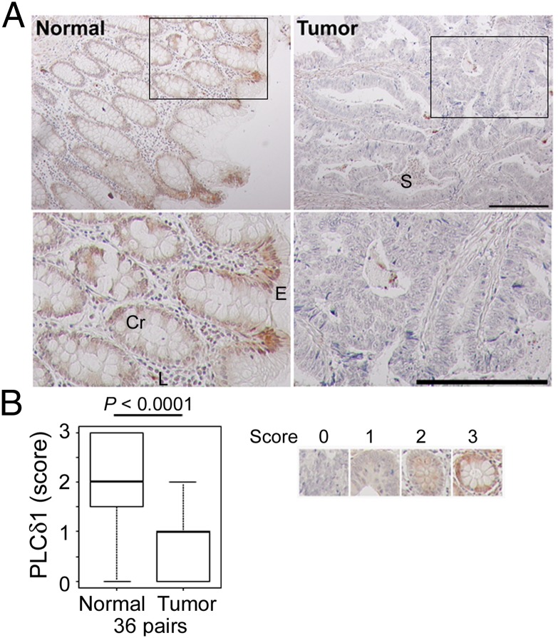
Fig. 1.
PLCδ1 was down-regulated in colon adenocarcinoma. (A) Immunohistochemistry with human colon carcinoma tissue arrays, which contain 36 matched normal and adenocarcinoma tissues, was performed with anti-PLCδ1 antibody. Insets in Upper are shown magnified in Lower. Cr, crypt; E, surface epithelium; L, lamina propria; S, stroma. (Scale bar: 200 µm.) (B) The expression level of PLCδ1 in each sample was scored. As shown in Right, scores of 0–3 indicate complete loss, mild staining, moderate staining, and marked staining, respectively. The scored PLCδ1 levels were assessed between normal and tumor samples (36 pairs) using the Wilcoxon signed rank test.

