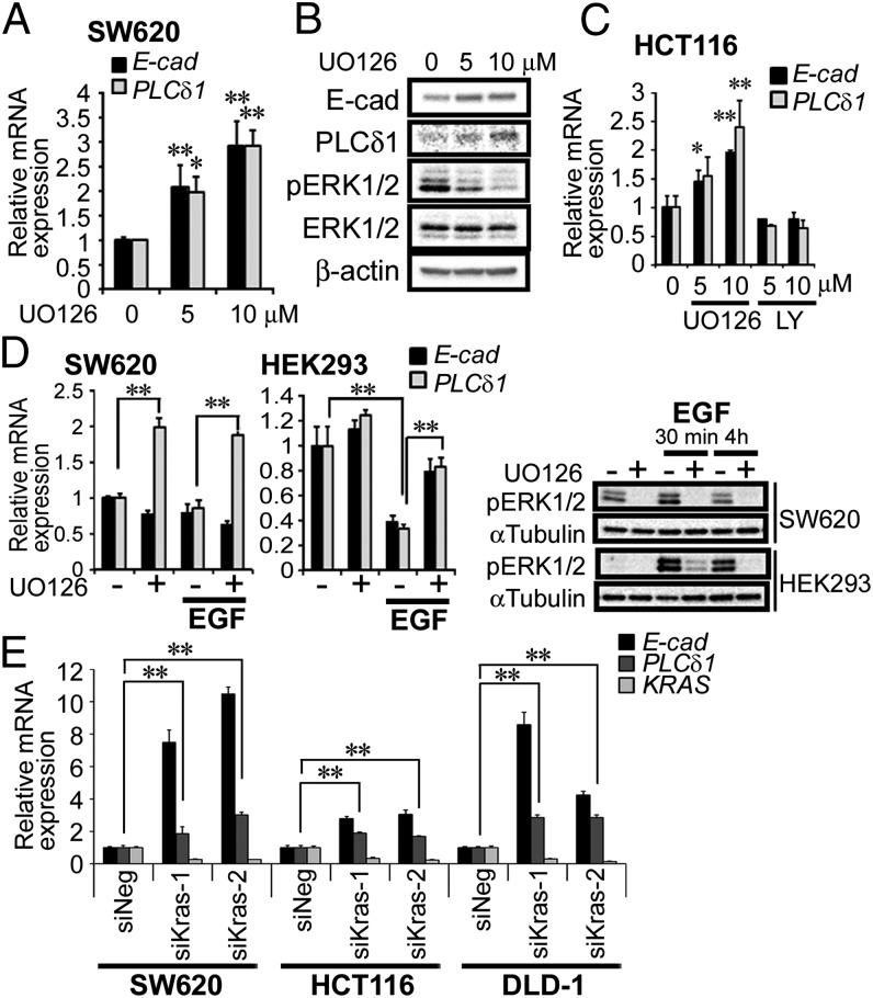
Fig. 6.
KRAS/MEK signaling suppressed the expression of PLCδ1. (A) SW620 cells were treated with the MEK inhibitor UO126 (5 or 10 µM) for 48 h. Cells were harvested, and the mRNA expression levels of PLCδ1, E-cadherin, and β-actin (as internal control) were determined by qRT-PCR analysis (n = 3). The relative expression levels of PLCδ1 and E-cadherin, normalized by β-actin, are shown. E-cad, E-cadherin. (B) SW620 cells treated as in A were assessed by Western blots with the indicated antibodies. (C) HCT116 cells were treated with UO126 (5 or 10 µM) or LY294002 (5 or 10 µM) for 24 h, and then, the mRNA expression levels were determined (n = 3). The relative expression levels of PLCδ1 and E-cadherin, normalized by β-actin, are shown. (D) SW620 and HEK293 cells were treated with UO126 (10 µM) or EGF (50 ng/mL) in FBS-free medium at indicated combinations for 4 h; then, cells were harvested, and the mRNA expression levels were determined (n = 3). The relative expression levels of PLCδ1 and E-cadherin, normalized by β-actin, are shown. (D, Right) Cells were also assessed by Western blots for the indicated proteins. (E) SW620, HCT116, and DLD-1 cells were transfected with negative control siRNA (siNeg) or siRNA targeting KRAS (siKras-1 or -2) for 48 h. The relative mRNA expression levels of PLCδ1, E-cadherin, and KRAS, normalized by β-actin, are shown (n = 3). Statistical analysis was performed using Tukey multiple comparison of means test. *P < 0.05; **P < 0.005.

