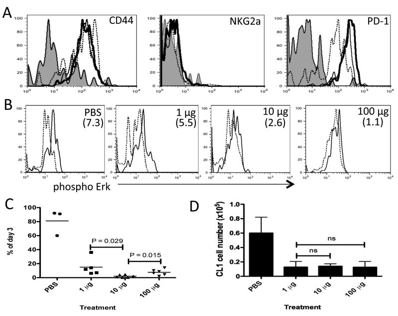
Figure 3. CD8+ T cells expressing a low affinity TCR for tolerizing antigen are not efficiently anergized.
B10.D2 mice containing donor Thy1.1+ CL1 CD8+ T cells were treated daily with PBS, 1, 10 or 100 μg of native Kd-HA peptide between days 0-2. (a) Day 3 flow cytometric analysis of donor CL1 cells in the spleen stained with antibody specific for indicated molecule. Lines; Shade –PBS, dashed – 1 μg, thin – 10 μg and thick – 100 μg treatments. (b) Day 3 ex vivo phosphorylation of Erk in CL1 cells re-stimulated with Kd-HA peptide (thin line) or unstimulated (dashed line). Treatment condition indicated in top right hand corner of plots and the mean MFI increase from unstimulated to stimulated cells in parenthesizes. (c) Survival of CL1 cells in the spleen. Expressed as the percentage of CL1 cells detected by FACS on day 6 compared to day 3 for each treatment condition. (d) On day 30 mice were challenge with PR8 influenza virus (I.P 500 HA units) and six days later donor CL1 cells were detected by FACS in the spleen. Each graph is representative of 3 experiments using age and sex matched mice. In the experiments depicted in c and d there were between 3 to 6 mice per treatment group.

