Figure 2.
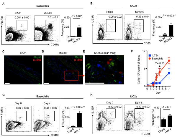
Basophils and ILC2s are enriched in murine AD-like skin lesions. (A) Skin basophils and (B) ILC2s in control vehicle (ethanol, EtOH) or MC903-treated wild-type (WT) mice. Cell frequencies are noted as a percentage of Lin− cells. (C) IF staining of Mcpt8+ basophils (green) and IL-33R+ ILC2s (red) cells in EtOH-treated and (D) MC903-treated Rag1−/− mice at low and (E) high magnification. DAPI+ cells are blue. Scale bars, 100 μm. All mice were treated topically daily for 7 days. (F) Skin basophils and ILC2s in MC903-treated WT mice on days 0, 2, 3, 4 and 7. (G) Skin basophils and (H) ILC2s in MC903-treated WT mice on days 0 and 4. Cell frequencies are noted as a percentage of Lin− cells. Data are representative of three independent experiments; n = 3 mice per group per experiment. Actual P-values are indicated.

