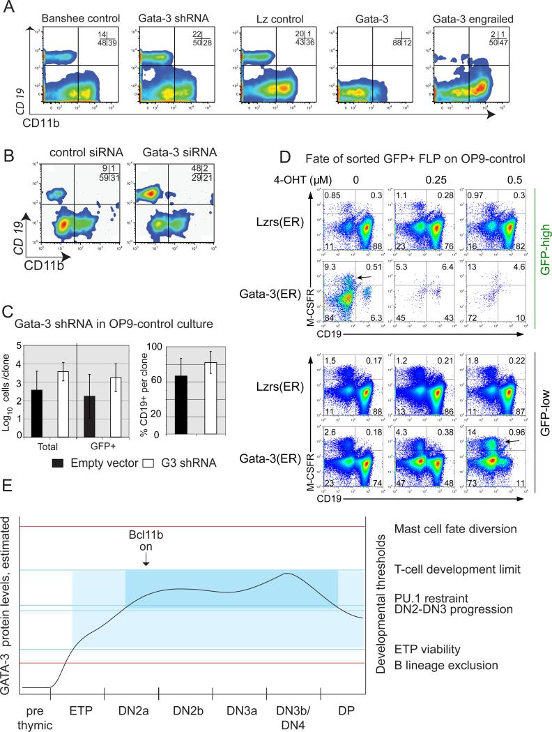
Figure 9. Expression of inducible GATA-3 and obligate repressor GATA-3 show distinct mechanisms for GATA-3 inhibition of B and myeloid development.
A. GATA-3 knockdown enhances B-cell emergence; GATA-3ENG expression blocks B-cell emergence from FLP. Bcl-2 transgenic FL precursors were infected in parallel with either G3-3W or Banshee control constructs, or with retroviral constructs overexpressing normal GATA-3 or an obligate repressor form of GATA-3 (Gata-3 Engrailed), or the corresponding vector control (Lz). Sorted FL precursors were then cultured 3.5 days with OP9 control stromal cells (B/myeloid conditions) before analysis (three independent experiments).
B. Loss of GATA-3 promotes B-cell emergence. FL precursors were nucleofected with siRNA specific for Gata3, then sorted and cultured 4-6 days under B/myeloid conditions on OP9 control stroma. Representative data shown (7/10 experiments using nucleofection or retroviral transduction of Gata-3 shRNA).
C. B-cell recovery from GATA-3 knockdown in clonal precursors. Single FL precursors transduced with Banshee or G3-3W were sorted into 96 well plates with OP9 control stroma and cultured for 12 days. Cell numbers for total cells/clone and GFP+ cells/clone are shown for G3-3W and control infected clones (left)(geomean ± s.d.). Percentages of each clone which were CD19+ are shown (right). At least as many B cells were generated from cells remaining GFP+ after transduction with 3W as with Ban; differences were not statistically significant.
D. Dose-dependent hierarchy of B-cell and myeloid inhibition by tamoxifen-enhanced GATA-3 [GATA3(ER)]. Fetal liver precursors were infected with GATA3(ER) or empty vector [Lz(ER)] and sorted the next day to purify GFP+ transductants. GFP+ cells were cultured for 6 days on OP9-control in the absence or presence of tamoxifen as indicated. Some CD45+ cells continued strong vector expression (GFP-high) while others downregulated it (GFP-low). These were analyzed for B (CD19+) and myeloid (CD115+) development (representative of two independent experiments). Concordant results were obtained in an additional experiment under different culture conditions (not shown).
E. Natural changes in GATA-3 levels cross distinct developmental dose thresholds. Schematic of effects shown here and in other work (3, 7, 11, 14, 44). Red lines: thresholds seen in absence of Notch signaling. Blue lines: thresholds seen in presence of Notch signaling. Light blue zone: permissive for T-lineage entry. Darker blue: permissive for full T-cell commitment.

