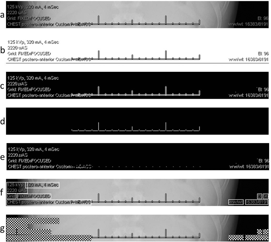
Fig. 4.
Intermediate and final results of the PHI detection and redaction processes. (a) The subregion of the original un-altered radiograph as described in Fig. 3. (b) The original image after application of the interpolation-OCR method, involving a brightness threshold filtering followed by bicubic interpolation. This image was tested with OCR algorithms without success. The threshold-redaction algorithm was as follows, with the step letter indicating the corresponding sub-figure: (c) The original image after application of a low-threshold filter. (d) The original image after application of a high-threshold filter. (e) An image obtained after performing a single pixel expansion of image 4d and subtract it from image 4c. (f) Determination of bounding boxes overlaid on the original image. (g) Redacted of alpha-numeric information inside the bounding boxes suing a checkerboard pattern applied to the original image.

