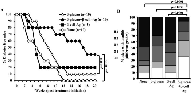
FIGURE 9. Treatment using β-glucan and β-cell-Ag resulted in better protection of NOD mice from hyperglycemia as compared to treatment with β-glucan or β-cell-Ag alone.
Twelve-week old euglycemic female NOD mice were left untreated (control) or treated with β-glucan (i.v.; on days 1, 3, 5, 13, 15, and 17 with 5 μg/mouse/day). Some groups of animals received β-cell-Ag on days 5 and 17. A) Mice were checked every week for hyperglycemia and blood glucose level of 250 mg/dl for two consecutive weeks was considered diabetic. Log-rank test was performed to compare hyperglycemia incidence in β-glucan and β-glucan plus β-cell-Ag treated groups with control and the p-value is shown on each graph. The group that received β-cell-Ag was also compared to control mice. B) One set of treated and control mice from parallel experiments were euthanized 4 weeks after the last injection. Pancreatic tissues were processed for H&E staining to evaluate insulitis as described above for Fig. 2. The islets with representative insulitis grades and percentages of islets with different grades of insulitis plotted as bar diagram are shown. Sections of pancreatic tissues from at least 5 mice/group were examined for insulitis and the insulitis score of at least 150 islets/group were plotted as bar diagram.

