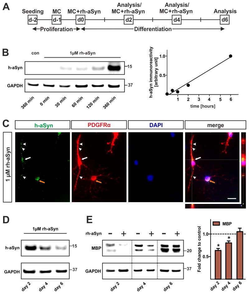
Fig. 6. Impaired maturation of primary OPCs upon uptake of rh-aSyn.
(A) Experimental paradigm to assess the effect of rh-aSyn on primary OPC maturation. OPCs were exposed to 1μM rh-aSyn 2 days after seeding prior to differentiation for additional 6 days. MC = medium change. (B) Western blot of primary OPCs lyzed at 0, 30, 60, 120, and 360 minutes upon rh-aSyn administration. Densitometry reveals that primary OPCs take up rh-aSyn in a time dependent manner. (C) Immunocytochemistry demonstrates the presence of intracellular rh-aSyn (green) in PDGFRα-positive primary OPCs (red) at day 2 upon exposure to rh-aSyn. Three-dimensional analysis using the z-stack module (AxioVision, ZEISS) verifies the presence of intracellular rh-aSyn rather than membranous attached rh-aSyn (merged picture). Note that bipolar PDGFRαhigh OPCs show h-aSyn immunoreactivity in the perinuclear cytoplasm (white arrow) as well as in processes (white arrowheads) whereas multipolar PDGFRαlow oligodendrocyte precursors only exhibit perinuclear h-aSyn signals (orange arrow). Scale bar: 20μm. (D) Representative Western blots (n=3) of whole cell lysates show a decline of the intracellular h-aSyn level within the course of differentiation. (E) Representative Western blot (n=3) of primary oligodendrocytes differentiated for 2, 4, and 6 days in the presence of rh-aSyn (1μM) shows the MBP expression pattern. Densitometric analysis reveals a significant reduction in MBP levels restricted to early stages (day 2 and day 4) of oligodendrocyte differentiation upon exposure to rh-aSyn.

