
Figure 2.
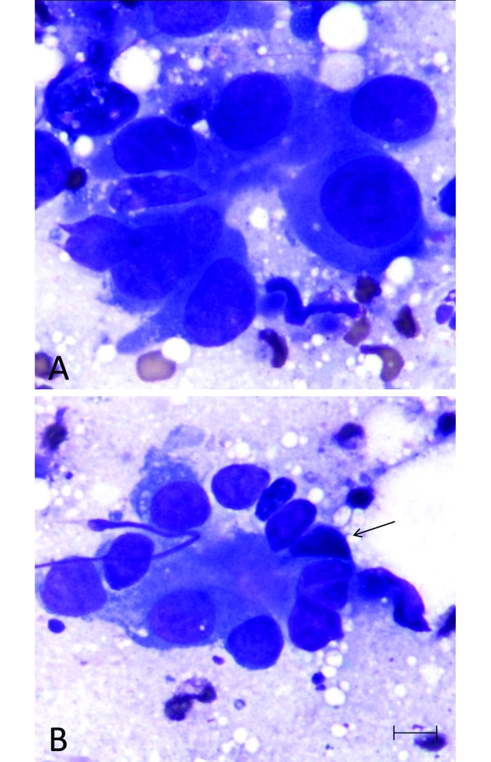
Fine-needle aspirate of the mammary tumor of case 1. (A, B) Clusters of polygonal epithelial cells arranged in crude acini are present with (arrow, B) occasional angular basal epithelial cells. Cells have distinct borders and variable amounts of basophilic vacuolated cytoplasm. Nuclei are round to oval to irregular, with finely stippled chromatin and 1 or 2 variably prominent dark-magenta nucleoli. Anisocytosis and anisokaryosis are moderate. Modified Giemsa stain; scale bar, 10 μm.