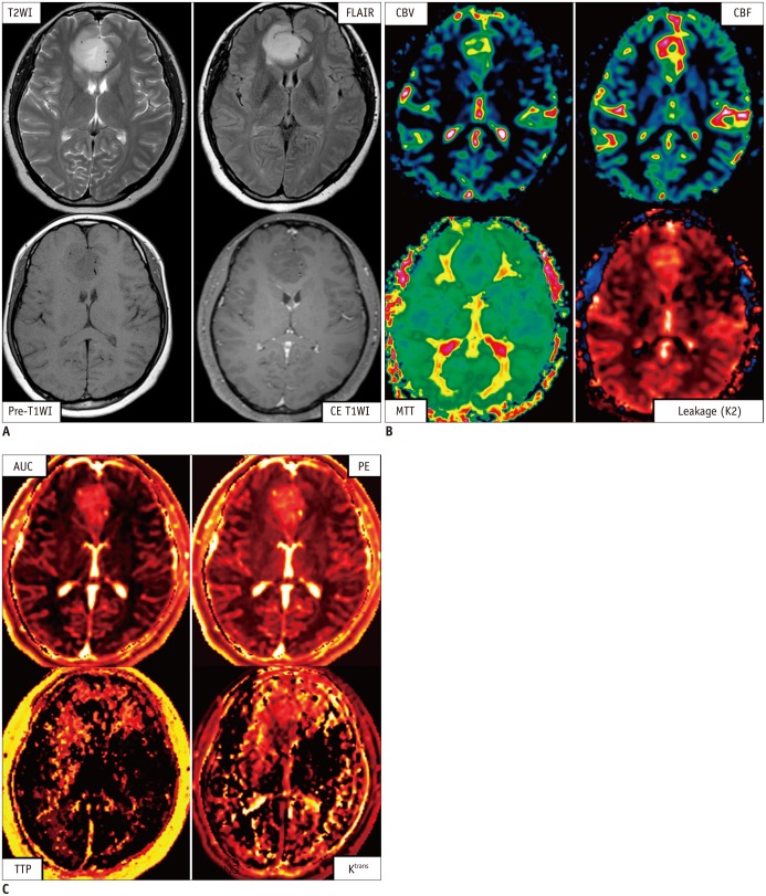
Fig. 4.
Case of clinical application of perfusion MRI methods in patient with brain tumor.
MR images and parameter maps (A) calculated from data of both dynamic susceptibility-contrast MRI (B), and dynamic contrast-enhanced MRI (C), obtained from patient who has abaplastic astrocytoma (World Health Organization grade III) in frontal lobe in brain. Brain-blood barrier is intact (CE T1WI), but tumor vascularity is increased. AUC = area under curve, CBF = cerebral blood flow, CBV = cerebral blood volume, CE T1WI = T1-weighted image after injecting contrast agent, FLAIR = fluid attenuated inversion recovery image, MTT = mean transit time, PE = peak enhancement, Pre-T1WI = T1-weighted image before injecting contrast agent, TTP = time-to-peak, T2WI = T2-weighted image

