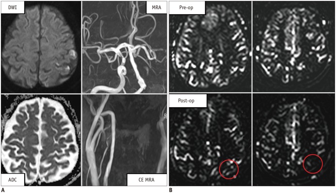
Fig. 5.
Case of clinical application of arterial spin-labeling MRI in patient with brain infarction.
Magnetic resonance images (A), and perfusion-weighted imaging (B), before (Pre-op) and after (Post-op) bypass surgery, in 59-year-old male with border zone infarction. A. Image shows DWI obtained with b-value of 1000 s/mm2, and corresponding ADC map, time-of-flight MRA, and CE MRA. High signal intensity on DWI at left side of brain indicates area with decreased diffusion, and MRA shows occlusion of middle cerebral artery. B. Image shows two slices of perfusion-weighted images before, and after bypass surgery. Slightly increased CBF is shown after bypass surgery. Only small amount of CBF is observed, because images were obtained immediately after bypass surgery. ADC = apparent diffusion coefficient, CBF = cerebral blood flow, CE MRA = magnetic resonance angiography with injecting contrast agent, DWI = diffusion-weighted imaging, MRA = magnetic resonance angiography with time-of-flight technique

