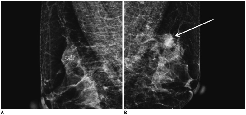
Fig. 4.
Example case of choice 4 in group practice reading.
Right mediolateral oblique (MLO) view (A) shows clustered fine pleomorphic calcifications in upper portion and left MLO view (B) shows round, obscured hyperdense mass (arrow) in upper portion. These were confirmed to be ductal carcinoma in situ in right breast and invasive ductal carcinoma in left breast.

