Figure 4.

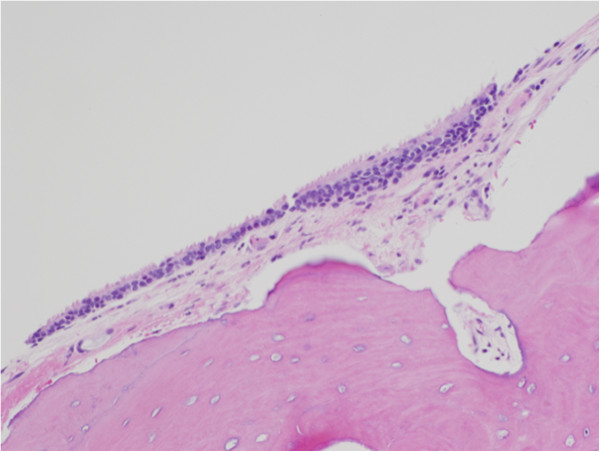
Histopathological section of middle ear osteoma showing the bone covering of cuboidal ciliated epithelium with a thin band of intervening fibrous tissue (stained with hematoxylin and eosin).

Histopathological section of middle ear osteoma showing the bone covering of cuboidal ciliated epithelium with a thin band of intervening fibrous tissue (stained with hematoxylin and eosin).