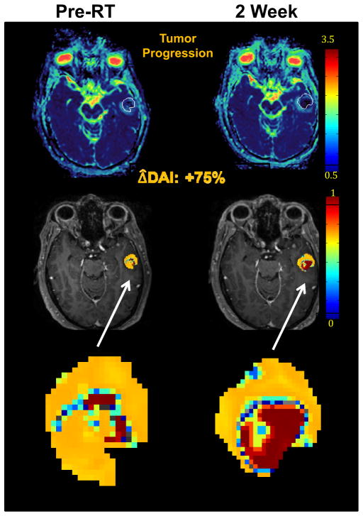
Fig 1.
Diffusion abnormality index (DAI) of a patient with a brain metastasis treated with whole brain radiation therapy. DW MRI was obtained both pre RT (left column) and at the end of RT (right column). Top row: color-coded ADC maps; middle row: DAI maps; bottom row: zoomed DAI maps of the tumor. The tumor volume showed no significant change from pre RT to the end of RT. However, the DAI increased by 75% from pre RT to end of RT, suggesting tumor progression which was confirmed by post-Gd T1-weighted MRI one month post RT.

