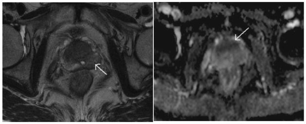
Fig 3.

A 72-year-old patient with a serum PSA level of 4.1 ng/mL, the T2W imaging (left panel) showed a hypointense area in the left peripheral zone (arrow); discrimination of cancer from BPH in the transition zone was difficult. The ADC map (right panel) depicted a cancer focus in the anterior prostate as a hypointense area (arrow). Pathologic results revealed a cancer focus well correlated with the finding of the ADC map, but no malignancy was found in the left peripheral zone. Reprinted with permission from Miao et al in Eur J Radiol 2007;61:297–302.
