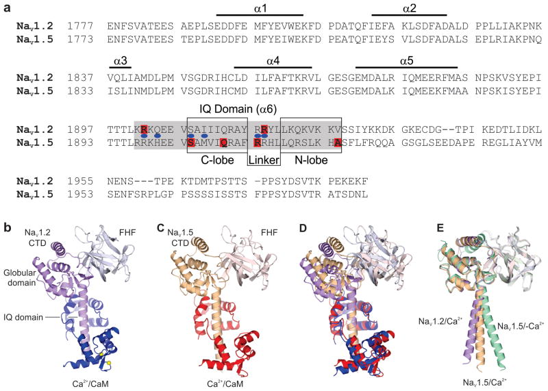
Figure 1. Overall architecture of the ternary complexes containing a NaV CTD, CaM, and a FHF.
(A) Sequence alignment of the NaV1.2 and NaV1.5 CTDs, with structural motifs and the IQ domain indicated. The interaction sites for the CaM N- and C-lobes and the CaM interlobular linker are also indicated. Human disease mutations in NaV1.2 and NaV1.5 clustering in the IQ domain are indicated in red; in blue are the homologous positions of disease mutations in NaV1.1. (B) The NaV1.2/Ca2+ structure containing the NaV1.2 CTD (purple), FGF13 (silver), and CaM (blue). (C) The NaV1.5/Ca2+ structure containing the NaV1.5 CTD (orange), FGF12 (pink), and CaM (red). (D) Overlay of the NaV1.2/Ca2+ and the NaV1.5/Ca2+ structures aligned to the IQ motifs. (E) Overlay of the NaV1.2/Ca2+ structure; the NaV1.5/Ca2+ structure; and the NaV1.5/-Ca2+ structure (Protein Data Bank accession code 4DCK), all aligned to the globular domain of the respective CTDs. For clarity, their respective CaM structures were omitted. This arrangement emphasizes the different angles between the globular domains and the IQ motifs among the three structures.

