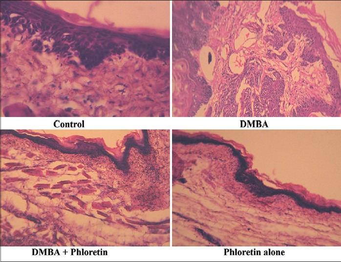
Figure 1.

Histopathological evaluation of DMBA induced hamster buccal pouch carcinogenesis. Microphotograph of control animals showing normal epithelium in buccal mucosa. Microphotograph of DMBA alone treated animals showing well differentiated squamous cell carcinoma exhibiting keratin pearls in the connective tissue. Microphotograph of DMBA+phloretin treated animals exhibiting mild hyperplasia and mild dysplasia. Microphotograph of phloretin alone treated animals showing normal epithelium in buccal mucosa
