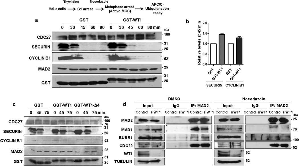
Figure 5. WT1 inhibits APC/C function.
(a) APC/C ubiquitination assay was performed with mitotic extracts derived from nocodazole-arrested HeLa cells in the presence of GST control or GST-WT1 (residues 245–297) for different time periods. The reaction was stopped at 0, 30, 45, 60, and 90 minutes and immunoblotting was performed with the indicated antibodies. The degradation of SECURIN and CYCLIN B1 was monitored for the indicated time points. (b) The band intensity at 45 min time point for SECURIN and CYCLIN B1 degradation in the presence of GST control or GST-WT1 from three independent experiments was quantified using Image J software and plotted graphically. Error bars are standard deviation from the mean. (c) APC/C ubiquitination assay was performed as in part A, with GST, GST-WT1 (residues 245–297) and GST-WT1Δ4 (Δ 288–295) at different time points and immunoblotted with the antibodies indicated. (d) WiT49 cells were transfected with either control or WT1 siRNA for 48 hours followed by treating the cells with either DMSO or nocodazole (60 ng/ml) for a further 12 hours. Whole cell extracts were prepared and MAD2 was immunoprecipitated then immunoblotted with anti-MAD1, anti-BUBR1, anti-CDC20 and anti-MAD2 antibodies. Input is 10% of the total whole cell extract used for immunoprecipitation.

