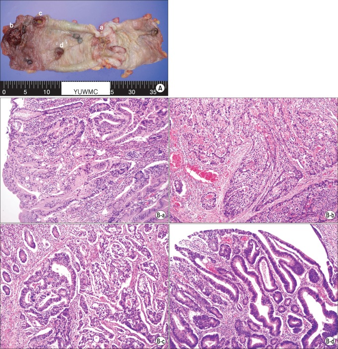
Fig. 2.
Histopathologic findings. (A) A total of four adenocarcinomas (a to d) are seen in the specimen. (B) The corresponding histopathologic examination (matched with tumor lesions, alphabetical order). There are one pT4 lesion (a), two pT3 lesions (b, c), and a carcinoma in situ lesion (d) (H&E, ×200).

