
Figure 4. ASP3026 suppresses NPM-ALK+ ALCL tumor cell growth in vivo.
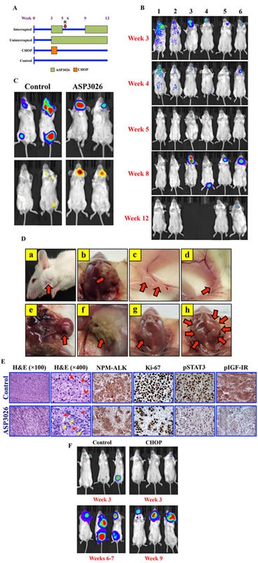
(A) C.B-17 SCID mice were randomized into the 4 illustrated treatment groups at week 3 after intravenous injection of Karpas 299 cells permanently expressing firefly luciferase. “R” denotes relapse after ASP3026 discontinuation. (B) Mice developed NPM-ALK+ ALCL tumors within approximately 3 weeks after injection, and were monitored weekly using bioluminescence imaging. The intensity of the signal is indicated by color (blue: low; green: intermediate; red: high tumor burden). Mice uninterruptedly treated with ASP3026 (examples shown in lanes 1 & 2) underwent complete remission with no relapse until study termination at week 13. Administration of ASP3026 was interrupted in 5 mice (examples in lanes 3-6) for 4 weeks. Notably, ALCL relapsed as early as 1 week after discontinuation of ASP3026. Two mice (lanes 3 & 4) had to be euthanized before the study period ended because of relapsed NPM-ALK+ ALCL with extensive tumor burden. At week 9, ASP3026 was re-administered in the other 3 mice (2 shown in lanes 5 & 6), and the NPM-ALK+ ALCL tumors substantially regressed, and the mice survived until termination of the study. (C) Bioluminescence imaging highlights the NPM-ALK+ ALCL tumors in control and ASP3026-treated mice (upper panels). Mice were injected intraperitoneally by the firefly luciferase prosubstrate VivoGlo Caspase-3/7 and bioluminescence imaging was performed. Tumors in control mice treated only with vehicle failed to demonstrate significant apoptosis signals (left lower panel). In contrast, intense signals consistent with marked in vivo lymphoma cell apoptosis were detected in mice treated with ASP3026 (right lower panel). (D) Examples of control mice showing lymphoma (red arrows) in cervical lymph nodes (a, exterior; b, dissected); inguinal lymph nodes (c, d); mesenteric lymph nodes (e); and axillary lymph nodes (f). Example of cervical lymph nodes is shown in a mouse treated interruptedly with ASP3026 (group I). After relapse, this mouse received ASP3026, survived, and was euthanized at the end of the study. A small cervical lymph node (red arrow) was detected at necropsy (g). This lymph node was processed and microscopically examined as shown in Figure 3E, lower panel. Example of cervical lymph nodes is shown after relapse in a mouse treated interruptedly with ASP3026. The lymph nodes comprised large, bulky tumor masses (red arrows) (h). (E) Histologic sections from cervical lymph node obtained from a control mouse treated with vehicle show effacement of the normal nodal architecture by solid sheets of lymphoma cells (H&E staining; original magnification: ×100). Higher magnification shows large cells with abundant cytoplasm, vesicular nuclei, and high mitotic rate with atypical mitotic figures [red arrowheads] (H&E; ×400). Immunohistochemical staining shows cytoplasmic and nuclear localization of NPM-ALK protein in the solid sheets of the lymphoma cells. Ki-67 is expressed in the vast majority of the lymphoma cells that diffusely infiltrate the lymph node. In addition, pSTAT3Y705 and pIGF-IRY1161 are highly expressed in almost all of the lymphoma cells. Also shown are histologic sections from a small post-treatment cervical lymph node collected at necropsy from a mouse treated with ASP3026 after relapse (I group, illustrated in Figure 3D, panel g). H&E staining shows small nests of ALCL cells separated by thick stromal tissue strands (×100). Higher magnification of the same lymph node further illustrates the small clusters of lymphoma cells with rare mitotic figures [yellow arrows] separated by thick stromal strands that include fibroblasts, small blood vessels, and admixed inflammatory cells [red arrows] (×400). Although NPM-ALK and Ki-67 were identified in residual ALCL cells, the numbers of these cells were much less pronounced after treatment. The expressions of pSTAT3 and pIGF-IR are substantially reduced in the lymph node sections from the ASP3026-treated mouse. Immunohistochemical staining photomicrographs are shown at an original magnification of ×400. (F) Control mice at week 3 (top left) and weeks 6-7 (bottom left) show remarkable progression of systemic NPM-ALK+ ALCL. Examples of mice from the CHOP-treatment group with NPM-ALK+ ALCL established at week 3 (top right). Although CHOP initially suppressed NPM-ALK+ ALCL tumor growth, lymphoma relapsed as illustrated at week 9 and the mice had to be euthanized because of heavy tumor burden (bottom right).