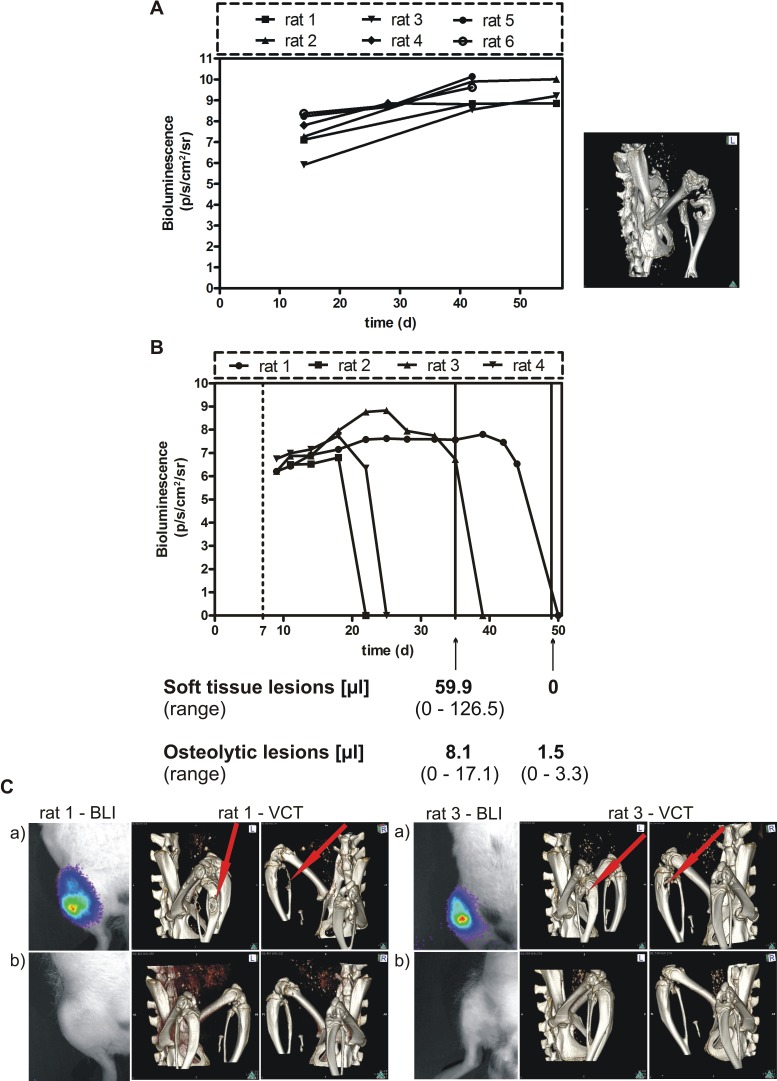
Figure 4. Effect of conditional BSP knockdown in vivo.
A. Spontaneous growth of the parental MDA-MB-231 cell line following injection into the femoral artery of nude rats. The tumor growth was estimated by BLI and the skeletal lesion monitored by VCT detection. B. BSP knockdown suppresses metastasis in vivo – bioluminescence imaging detection. B3 cells were inoculated into nude rats, which received doxycycline for 1 week (dashed line) and were exposed to miRNA treatment for up to 6 weeks; the time after tumor cell inoculation (days) is given on the x axis; bioluminescence is measured in photons/second/cm2/steradian, the volume of soft tissue and osteolytic lesions is indicated after 4 and 6 weeks of miRNA treatment (solid lines); C. The upper BL images (a) show the status after 4 weeks, the lower images (b) – that after 6 weeks (absence of light emission indicates absence of tumor, confirmed by histopathology); VCT scans performed after 4 weeks (upper panels (a), middle and right images) and after 6 weeks (lower panels (b), middle and right images) revealed complete remission of osteolytic lesions.

