
Fig 1. Hypoxia Primed Breast Cancer Cells Chemoattract Macrophages.
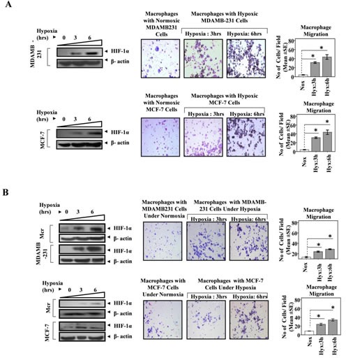
(A) MDA-MB-231 and MCF-7 breast cancer cells cultured in lower well of modified boyden chamber were exposed to hypoxic environment for 3and 6 hrs. THP-1 derived macrophages previously cultured on PET cell culture inserts were introduced in upper well. Extent of macrophage migration was evaluated after maintaining the co-cultures for further 24 hrs under standard cell culture conditions. (B) Breast cancer cells (MDA-MB-231 and MCF-7) and Macrophage (THP-1 derived macrophages) co-cultures were exposed to hypoxic environment for 3and 6 hrs. Extent of macrophage migration was evaluated after maintaining the co-cultures for further 24 hrs under standard cell culture conditions. Representative western blot data showing hypoxia mediated upregulation of HIF1-α in MDA-MB-231, MCF-7 breast cancer cells and THP-1 derived macrophages as an indication of hypoxic stress. Photomicrographs (100X) depict macrophage chemotaxic towards breast cancer cells as evaluated through Geimsa staining of migrated macrophages. Quantification of macrophage chemotaxis was done by DAPI staining of migrated macrophages followed by counting of nuclei in five different fields of three replica wells. Data presented as Mean±SEM; n=5; Symbols indicate statistical significance at p < 0.05 (*).