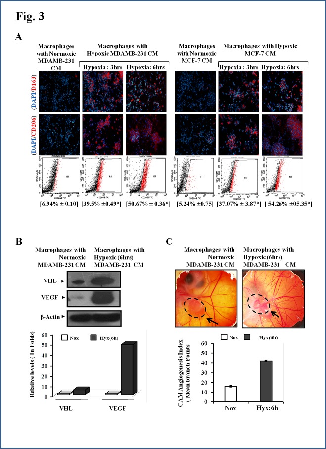
Fig.3. Enhanced M2-polarization of Macrophage with Potentiation of Pro-angiogenic Function by Hypoxic Breast Cancers Cells through the Release of Soluble Mediators.
THP-1 derived macrophages were incubated with conditioned media from hypoxia primed (3 and 6hrs) breast cancer cells (MDA-MB-231 and MCF-7) CM for 24 hrs, followed by phenotype evaluation using immunocytochemistry and flow cytometry. (A) Representative photomicrographs and flow cytometry data depicting enhanced M2-polarization of THP-1 derived macrophages in presence of hypoxia primed breast cancer cells conditioned media as measured through immunocytochemistry and flow cytometry analysis using Alexa fluor 555 or FITC conjugated anti-CD206 antibody respectively. Values in parenthesis represent mean ± SEM (n=3) of % M2-macrophage count obtained during flow cytometric analysis of three independent experiments. (B) Representative qualitative and quantitative western blot data showing hypoxia primed breast cancer cells conditioned media induced upregulation of key angiogenic regulators viz. Von Hippal-Lindau protein (VHL) and Vascular Endothelial Growth Factor (VEGF) with in macrophages. (C) Representative CAM assay stereozoom micrograph and CAM Angiogenesis index as a measure of angiogenic potential of macrophages incubated with normoxic or hypoxic breast cancer cells CM. Dotted circle represents gelatin sponge graft site.

