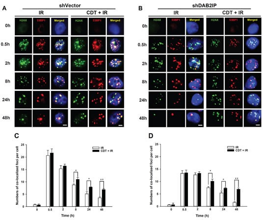
Figure 6. CDT enhances IR-induced DSB in DAB2IP-deficient PCa cells.

(A) shVector or (B) shDAB2IP cells were untreated (0 h), exposed to IR (2 Gy) alone, or treated with a combination of CDT (50 nM) and IR (2 Gy) for the indicated time and immunostained for phospho-γ-H2AX (green) and 53BP1 (red) foci. The number of co-localized foci (phospho-γ-H2AX and 53BP1) was determined for each time point in (C) shVector and (D) shDAB2IP cells. The remaining merged foci in the nuclei were counted in 3 independent experiments (50 nuclei each). Statistical significance was evaluated using Student's t-test (*, P < 0.05; **, P < 0.01).
