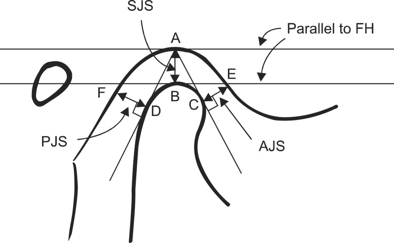
Fig. 2.
Tracing of temporomandibular joint tomogram to evaluate superior joint space (SJS), anterior joint space (AJS), and posterior joint space (PJS). (FH: Frankfort horizontal, A: the most superior point of the glenoid fossa, B: the most superior point of the condyle, C: tangent to the anterior surface of condyle from point A, D: tangent to the posterior surface of condyle from point A, E: intersection point perpendicular to A-C line from point C and anterior slope of the glenoid fossa, F: intersection point between the point perpendicular to A-D line from point D and the posterior slope of glenoid fossa)
Husanov Zafar et al: Positional change of the condyle after orthodontic-orthognathic surgical treatment: is there a relationship to skeletal relapse? J Korean Assoc Oral Maxillofac Surg 2014

