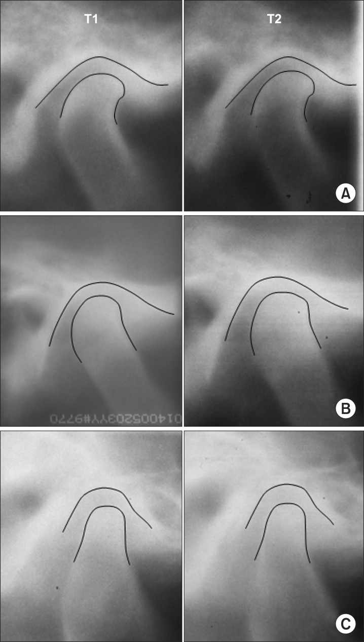
Fig. 3.
Temporomandibular joint tomograms at pretreatment (T1) and posttreatment (T2) showing three patterns of positional changes of the condyle. A. C-C pattern (unchanged). B. C-A pattern (forward movement). C. A-C pattern (backward movement). Refer to Table 3 for the definitions of patterns.
Husanov Zafar et al: Positional change of the condyle after orthodontic-orthognathic surgical treatment: is there a relationship to skeletal relapse? J Korean Assoc Oral Maxillofac Surg 2014

