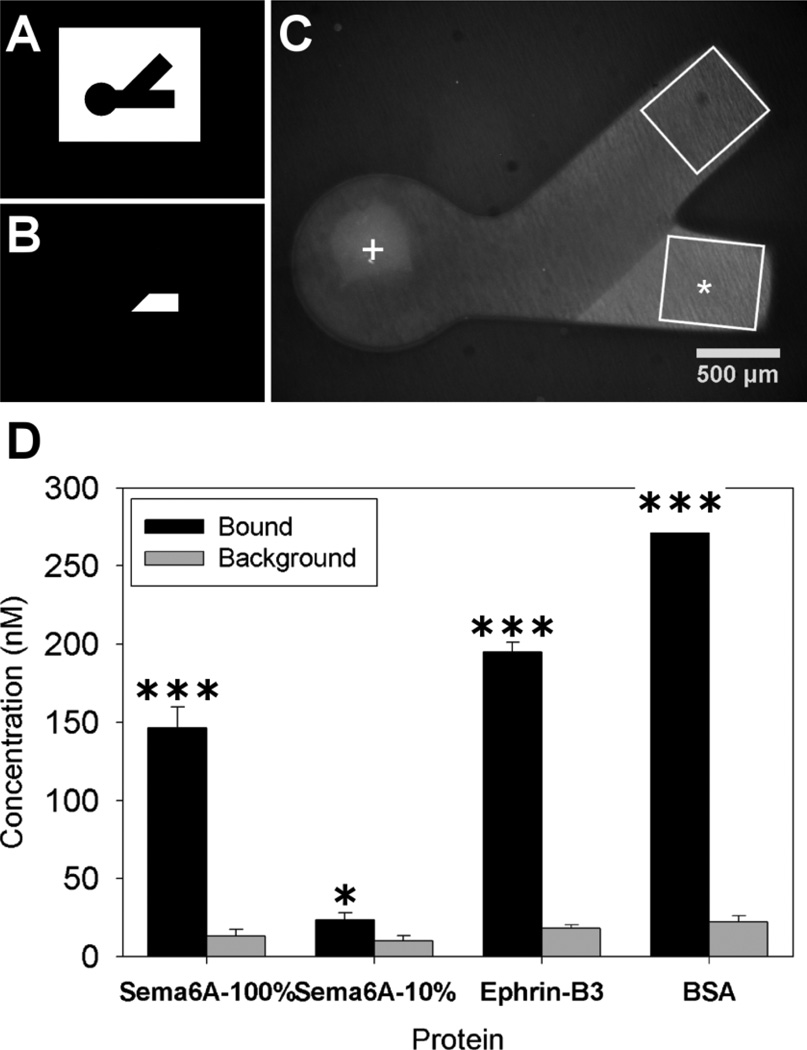
Figure 2.
Polymerization and protein binding for immobilized choice point models. Photomasks used for polymerization of PEG (A) and for spatial immobilization of proteins (B). Dual hydrogel construct used for immobilized protein experiments (C) with protein binding region indicated with *(Sema6A), white box approximating immunofluorescent ROI and + marks DRG explant location. The average concentration of immobilized protein in agarose (D) for the irradiated channel (bound) along with the average amount of protein seen in the non-irradiated channel (background) was plotted for each of the separate proteins used (n=10). The amount bound in irradiated regions was significantly higher than background fluorescence in non-irradiated regions for all bound proteins (student’s t-test, * p < 0.05 and *** p < 0.001).

