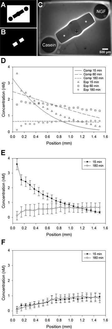
Figure 3.
Polymerization and protein diffusion for gradient choice point models. Photomasks used for polymerization of PEG (A) and for spatial immobilization of proteins (B). Dual hydrogel construct used for soluble gradient experiments (C) with protein binding region indicated with *(BSA), and + marks DRG explant location. D, Computational and experimental concentration profiles of soluble NGF diffusion through CNBC-agarose regions, demonstrating that the initial gradient formation and behavior through time was as expected. E, Experimental concentration profiles demonstrating gradient upon establishment and before refilling. F, NGF concentration profile in opposite channel was minimal and in the opposite direction. Concentration values were determined along center of channel with position 0.0 mm corresponding to the end of the channel nearest the soluble NGF reservoir (D,E) or soluble casein reservoir (F); DRG located at ~1.5 mm (n=3).

