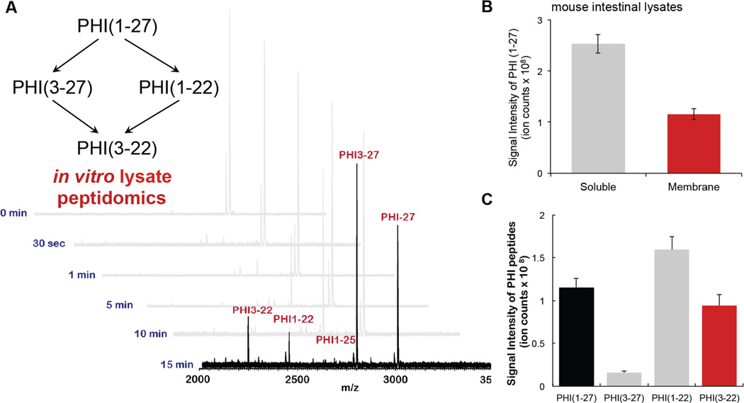
Figure 2.
Lysate peptidomics. A. MALDI profiles of in vitro assay using gut membrane lysates with gut peptide PHI1–27 for optimization of reaction time. B. PHI1–27 (100 µM) was incubated with the soluble (1 mg/mL) or membrane (1 mg/mL) fraction of the intestinal proteome (lysate) for 15 min. The sample was then quenched and analyzed by LC–MS to quantify PHI1–27 levels. A majority of the PHI-degrading activity was found in the membrane fraction, as evidenced by greatly diminished PHI1–27 levels after 15 min in the membrane fraction, and largely unchanged levels after exposure to the soluble fraction. C. PHI1–27 proteolytic fragments after incubation with intestinal membrane lysates.

