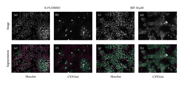
Figure 6.

Segmentation of HepaRG cells for CYP3A4 expression using IN Cell Workstation software. This figure illustrates image analysis software tracing specified features of interest in representative images of HepaRG cells seeded at 50,000 cells per well. Upper panels (a–d) show staining images while lower panels (e–h) show image segmentation via HCA image analysis algorithms. Nuclear segmentation is shown by blue outlines in (e–h); cells expressing CYP3A4 are shown by green outlines; cells not expressing CYP3A4 are shown by red outlines. Hoechst nuclear stain is used to identify and count all cells in the images (a + e; c + g). Effects of 10 μM rifampicin upon CYP3A4 expression versus vehicle control can be seen by comparing (d) with (b; h) and (f) shows the segmentation of CYP3A4 staining which enables subsequent quantitation.
