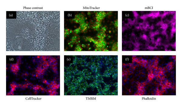
Figure 9.

Labeling of HepaRG with cell function and structure dyes. Images shown represent HepaRG cells plated on 96-well collagen coated plates in growth media at 50,000 cells per well and cultured for 3 days. Images shown represent label-free phase contrast imaging of live cells (a); live cells stained with MitoTracker Green FM (b); live cells stained with Monochlorobimane (mBCI) (c); live cells stained with CellTracker Red CMTPX (d); live cells stained with Tetramethylrhodamine, Methyl Ester, and Perchlorate (TMRM) (e); and fixed cells stained with Alexa Fluor 568 Phalloidin (f).
