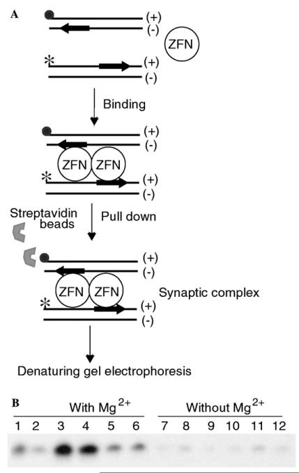
Fig. 3.
Biotin pull-down assay. (A) Schematic representation of the biotin pull-down assay. The filled circle represents the biotin tag and the star represents the 32P-label. The substrate encodes the ΔQNK site near the 3′ end of the (+) strand. (B) PAGE profile of the biotin pull-down assay. The biotin-tagged DNA was recovered from the reaction using streptavidin coated magnetic beads. The 32P-labeled DNA from the complex was analyzed by using PAGE. Lanes 1–6 with Mg2+ions; 1, control IVTT (without ZFN plasmid) + specific DNA; 2, control IVTT + non-specific DNA; 3, ΔQNK-FN (1 μl) + specific DNA; 4, ΔQNK-FN (4 μl) + specific DNA; 5, ΔQNK-FN (1 μl) + non-specific DNA; 6, ΔQNK-FN (4 μl) + non-specific DNA; and 7–12, same as lanes 1–6, but without Mg2+ions.

