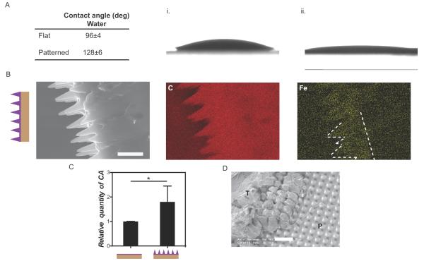
Figure 2.
Characterization of CA coated surfaces. A) Contact angles of water and CA on flat and patterned PCL samples. B) Lower CA spreadability was observed on flat (i) than on (ii) patterned substrates. C) Cross-section of CA spin-coated films on patterned PCL. Chemical mapping of IO nanoparticles encapsulated within CA demonstrated that CA covered the patterned area. Scale bar 10 μm. D) Quantification of CA on flat and patterned spin-coated PCL patches through ICP-AES (n=5 per condition). The CA amounts on spin-coated flat substrates were 13 ±2 times less than 5μL (non spin-coated) CA coated substrates (data not shown). E) SEM image of patterned PDMS after adhesion testing against ex vivo intestine tissue. Tissue residue (T) was visible on the patch surface and was interlocked with the surface topography (P). Scale bar 20 μm.

