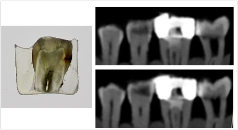
Figure 3.
Microscopic image of a section of a pre-molar tooth and the corresponding CBCT scans. Microscopic examination reveals the presence of enamel and dentine caries on distal side. CBCT examination suggests enamel and dentine caries on distal side but the extent of demineralization is more severe in comparison with histological view.

