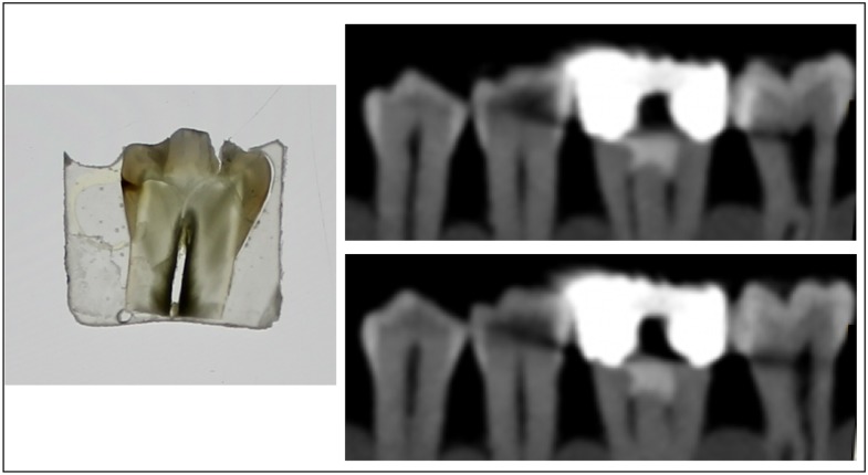
Figure 4.
Microscopic image of a section of a pre-molar tooth and the corresponding CBCT scans. Microscopic examination reveals the presence of enamel caries on the mesial side. CBCT examination is severely compromised by the presence of diagonally shaped artefact that makes diagnosis of enamel demineralization very uncertain.

