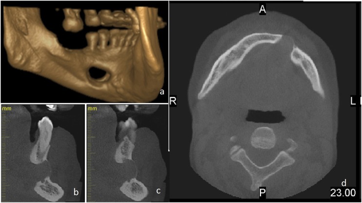
Figure 2.
Images taken from the CBCT examination. (a) Three-dimensional rendering of the mandible. The defect seems to have eroded the buccal cortex because of the burn out effect. (b, c) Cross-sectional images of the defect area showing the relationship of the inferior alveolar nerve and the apices of the teeth to the defect. (d) Axial image showing the buccal expansion caused by the defect. A, anterior; L, left; P, posterior; R, right.

