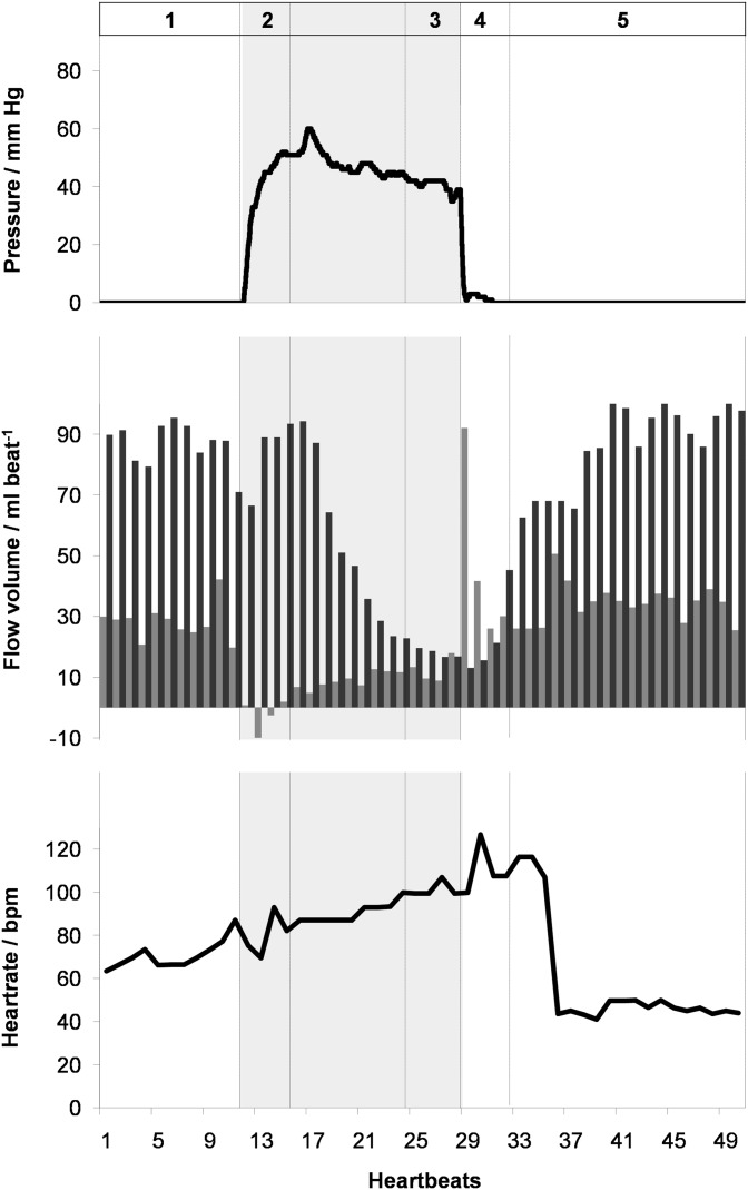
Figure 4.
(Top) pressure, (middle) flow volume in the ascending aorta (black) and superior vena cava (grey), and (bottom) heart rate as a function of time during the Valsalva manoeuvre (shaded) in a 24-year-old male. Real-time phase-contrast flow MRI was performed for 40 s corresponding to 50 successive heartbeats. 1, normal breathing; 2, early Valsalva; 3, late Valsalva; 4, early recovery; and 5, late recovery. bpm, beats per minute.

