
Figure 7.
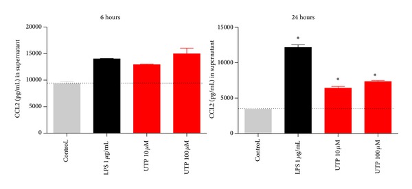
IFN/LPS differentiated THP-1 macrophages display increased CCL2 production to LPS or UTP. THP-1 cells were plated in RPMI media containing 10% serum and stimulated with IFN-γ and LPS for 48 hours. Media were removed and cells were challenged with either media alone (control), LPS (1 μg/mL), UTP (10 μM), or UTP (100 μM) for 4 hours. Mean raw data is plotted in pg/mL ± SEM. Standard curves were performed for each ELISA experiment with fits of r > 0.95.