
Figure 4.
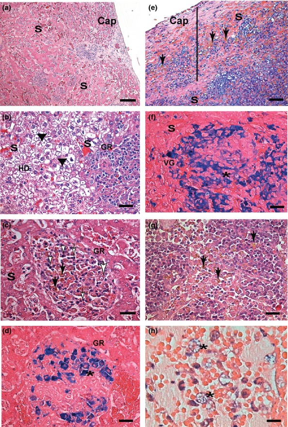
Liver, spleen and lymph node sections of symptomatic dogs naturally infected with L. infantum. Liver (a, b, c, d): (a) Note congestion of sinusoid vessels (S). Inflammation and capsule thickness (Cap). (b) Intense hydropic degeneration (HD), (black arrow heads) granuloma (GR) and congestion. (c) In the centre of the micrograph is an intralobular granuloma (GR) comprising epithelioid cells with an elongated or oval nucleus (large white arrow), plasma cells (white arrow heads) and lymphocytes (white small arrows). Haemosiderin deposition as circular brown structures can be noted (black arrows). (d) Perls' Prussian blue staining confirmed haemosiderin deposition inside macrophages of granulomas (*) where it has been visualized as homogeneous circular blue structures. (e, f) Spleen sections: (e) Note capsule (Cap) thickening (black bar) and congestion of the red pulp sinusoid vessels (S). Haemosiderin deposition as brown (black arrows). (f) Perls' Prussian blue staining confirming Virchow's granuloma (VG) haemosiderin deposition inside cells of the red pulp (macrophages*) visualized as homogeneous circular blue structures. (g, h) Lymph node sections: (g) Haemosiderin in macrophages (black arrows) of medullar and sinus cords. (h) Perls' Prussian blue staining confirming haemosiderin deposition inside foam macrophages (*). H&E (a, b, c, e and g); Perls' Prussia blue. (d, f, h); a, e (Bar = 32 μm), and b, c, d, f, g, h (Bar = 16 μm).