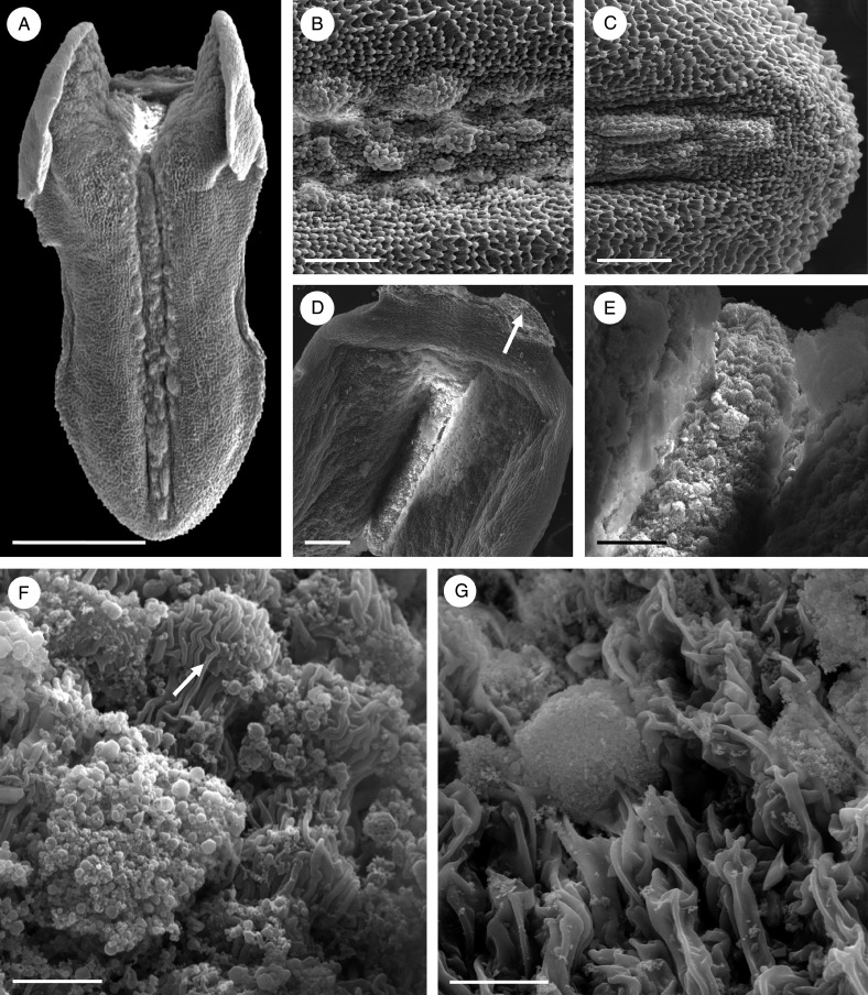
Fig. 10.
Bulbophyllum morphologorum – SEM. (A–C) Adaxial view of the labellum showing the presence of verrucae along the entire length of the median longitudinal groove. (D) Base of the labellum showing articulation (arrow) with the column-foot. (E) Basal part of the labellum coated with copious, amorphous secretion. (F) Detail of secretion and possible osmophore (arrow) with striate cuticle. (G) Squamous adaxial surface formed of palisade-like cells with a deeply striate cuticle and a globule of secretion. Scale bars = 1 mm, 200 μm, 200 μm, 200 μm, 50 μm, 10 μm and 20 μm, respectively.

