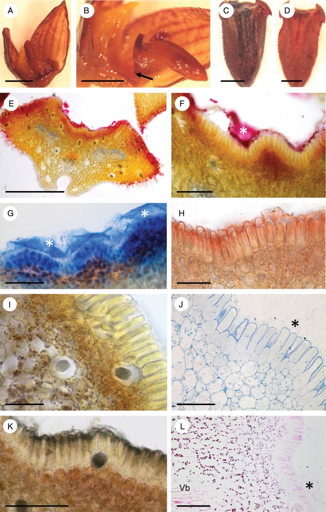
Fig. 6.
Bulbophyllum careyanum – LM. (A, B) Lateral view of the labellum showing articulation (arrow) to the column-foot. In (A) the rocking labellum is in the upright position and adpressed to the column, whereas in (B) it is in its usual lowered position. (C) Adaxial view of the labellum with the median longitudinal groove selectively stained for protein with Coomassie brilliant blue. (D) Adaxial view of the labellum with the median longitudinal groove selectively stained for mucilage with ruthenium red. (E, F) Transverse section of the labellum stained with ruthenium red. Note the stained secreted material (asterisk) coating the palisade-like epidermal cells, and the cuticular blister. (G) Section of labellum selectively stained for protein with Coomassie brilliant blue. The secreted material (asterisks) that has accumulated beneath the detached cuticle has stained strongly for protein. (H) Transverse section of the labellum showing the epidermis with the cuticle stained red following treatment with Sudan III, and small sub-epidermal cells with numerous plastids. (I) Section stained with IKI revealing the presence of starch in the sub-epidermal tissue and ground parenchyma, but its general absence from epidermal cells. Note also the presence of idioblasts with raphides. (J) Section of palisade-like epidermal cells and sub-epidermal tissue stained with MB/AII. Note the granular appearance of the secretion coating the epidermal cells and the cuticular blister (asterisk). (K) Control (unstained) section. (L) Palisade-like epidermal cells and parenchymatous tissues following PAS staining to show the distribution of starch. Note also the cuticular blister (asterisk). Scale bars = 2 mm, 2 mm, 0·5 mm, 0·5 mm, 500 μm, 100 μm, 100 μm, 50 μm, 50 μm, 50 μm, 100 μm and 100 μm, respectively. Vb, vascular bundle.

