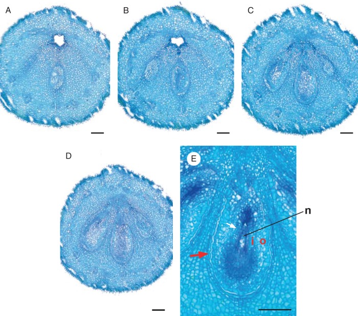
Fig. 5.
Emmotum harleyi. Transverse microtome section series of gynoecium at anthesis (A–D) and longitudinal section of ovule (E). (A) Level of apical septum (corresponding to Fig. 6H). (B) Level immediately below apical septum (corresponding to Fig. 6I). (C) Lowermost level of symplicate zone (corresponding to Fig. 6J). (D) Uppermost level of synascidiate zone (corresponding to Fig. 6 K). (E) Longitudinal section of bitegmic ovule. Signatures: o, outer integument; i, inner integument; n, nucellus; arrow, undulate contiguous surfaces of ovule and locular wall. Scale bars = 0·1 mm.

