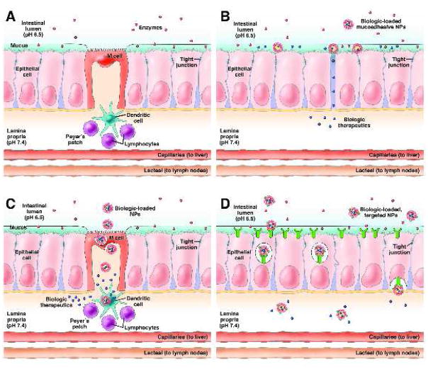
Figure 1.
(A) Schematic illustration of the barriers to oral biologic delivery presented by the GI tract, including acidic pH, enzymatic degradation, a mucus layer above the epithelial cells, the epithelial monolayer with limited permeability due to tight junctions, and immune cells associated with the M cells. (B) Mucoadhesive materials adhere to the mucus layer and release drug at high concentrations near the epithelial cells as well as reversibly open tight junctions to allow for biologic transport across the epithelium through the paracellular pathway. (C) The M cell transcytosis pathway is an approach used to enable NPs to cross the epithelium, but underlying dendritic cells associated with the Peyer’s Patches may endocytose NPs in the lamina propria. (D) Receptor-mediated transcytosis pathways such as the vitamin B12 receptor and FcRn can be targeted by NPs to enable transport across the epithelium and release into the lamina propria.

