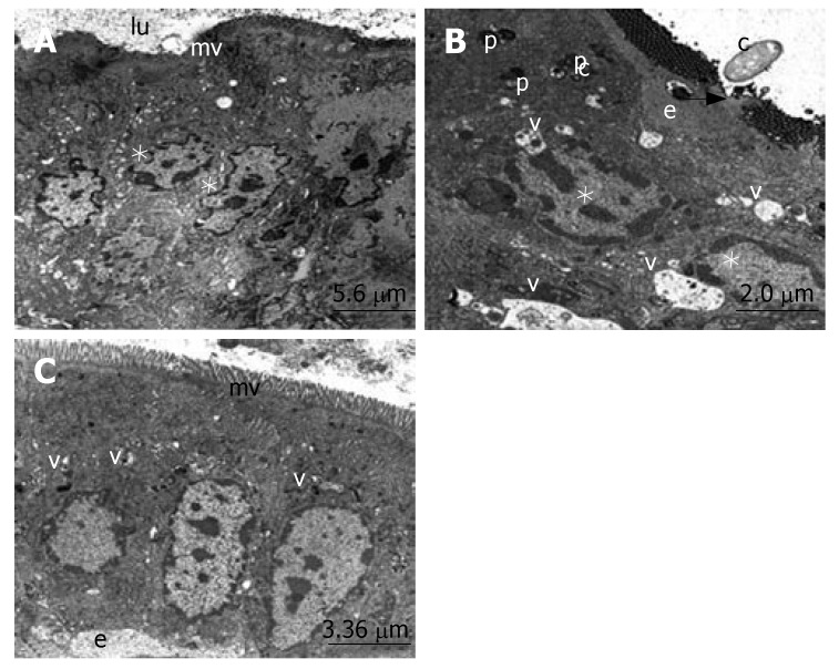
Figure 2.
These transmission electron microscope (TEM) micrographs illustrate the main ultrastructural features of enterocytes, and the absorptive cells of the ileum. A: The regular structure of microvilli (mv), lumen (lu) and the nuclei of the enterocytes (*). B: The subepithelial edema (e), phagosomes (p), vacuoles (v), desquamation of epithelial tissue (arrow), nuclei of enterocytes (*) and candida (c). C: The regular structure of microvilli (mv), small vacuoles (v) and subepithelial edema (e).

