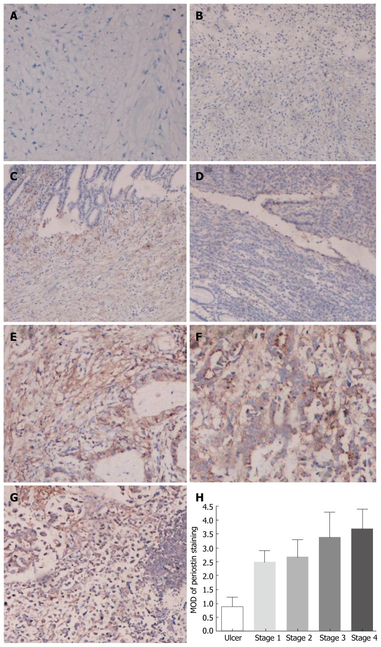
Figure 3.
Immunohistochemical analysis of periostin expression in normal gastric tissues, benign gastric ulcers, gastric cancer tissues, as well as lymphoid metastasis from gastric cancer. The tissue sections were immunostained with a polyclonal antibody. The positive staining for periostin protein is shown with a brown color. All sections were counterstained with hematoxylin showing a blue color. (A) Negative control; (B) benign gastric ulcer; (C) stageIgastric cancer; (D) stageIIgastric cancer; (E) stage III gastric cancer; (F) stage IV gastric cancer; (G) lymph node metastasis. The average MOD of periostin staining from stageI-IV gastric cancer was significantly higher than that from normal gastric tissues in each group (H) (P < 0.05).

