
Fig 4.
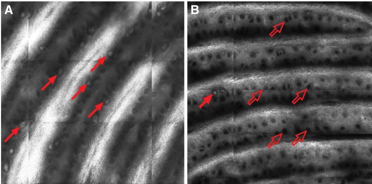
Representative Meissner's corpuscle (MC) scan images. Representative (A) healthy volunteer and (B) patient images of 1.0 × 1.0-mm in vivo laser reflectance confocal micrograph. In volunteer image, numerous MCs can be seen as white orb-shaped structures sitting at base of dermal papillae (dark bands). Several MCs are indicated by solid red arrows. One MC is visible in patient image, whereas numerous empty MC pits are visible, as indicated by open red arrows.