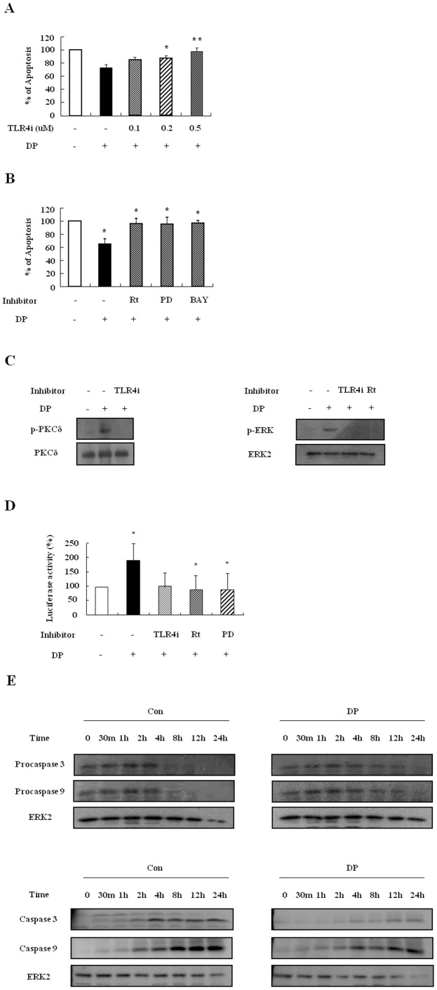
Figure 6. DP triggers suppression of constitutive apoptosis of AR neutrophils via activation of the TLR4/PKCδ/ERK/NF-κB pathway.
(A–B) AR neutrophils (at least n = 3) were pre-treated for 1 h with and without TLR4i in the indicated concentration (A), with and without 5 µM rottlerin (Rt), 20 µM PD98059 (PD), and 10 µM BAY-11-7085 (BAY) (B. Cells were incubated for 24 h in the presence and absence of DP (10 µg/ml). Apoptosis was analyzed by measuring the binding of annexin V-FITC and PI. Data are presented relative to the control, which was set at 100% as the means ± SD. *p<0.05 and **p<0.01 indicate a significant difference between the control and DP-treated groups or between the DP-treated and inhibitor-treated groups. (C) AR blood neutrophils were pre-treated for 1 h with and without 1 µM TLR4i or 5 µM rottlerin (Rt) and incubated with DP (10 µg/ml) for 5 min or 30 min. Harvested cells were lysed, and the phospho-PKCδ (p-PKCδ) (left panel) and phospho-ERK (p-ERK) (right panel) in the lysates were detected by Western blotting. (D) AR blood neutrophils were pre-treated for 1 h with and without 1 µM TLR4i, 5 µM rottlerin (Rt), or 20 µM PD98059 (PD) and then incubated with DP (10 µg/ml) for 8 h. After harvested cells were lysed, NF-κB in the lysates was detected by luciferase assay. Data are presented relative to the control, which was set at 100% as the means ± SD. *p<0.05 indicates a significant difference between the control and DP-treated groups or between the DP-treated and inhibitor-treated groups. (E) AR blood neutrophils were incubated with 10 µg/ml of DP for the indicated time. Procaspase 3, procaspase 9 (upper panel), caspase 3 and caspase 9 (lower panel) were detected by Western blotting. The membrane was stripped and reprobed with anti-ERK2 antibodies as an internal control.

