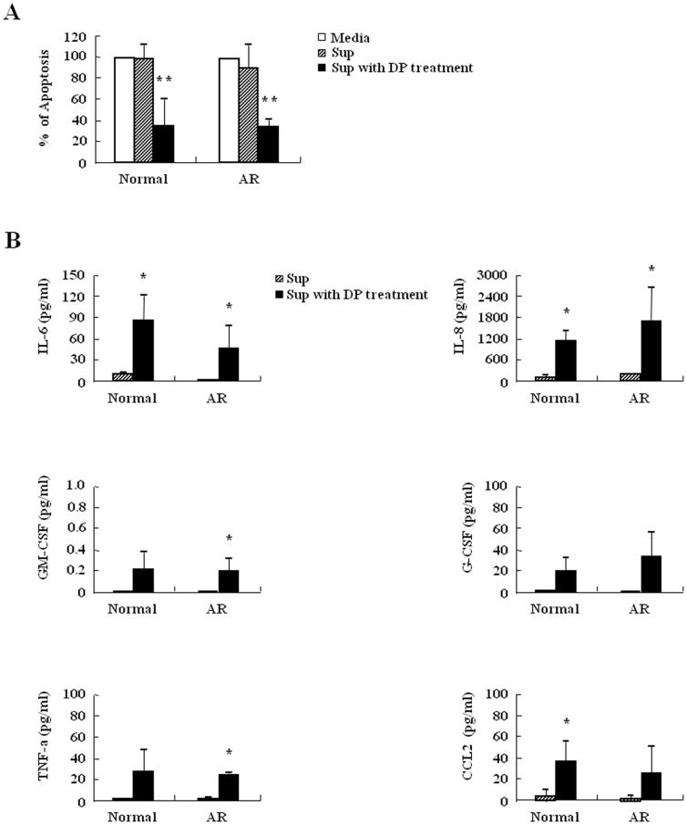
Figure 7. Inhibition of neutrophil apoptosis by DP is associated with molecules released by DP.
(A) Neutrophils from peripheral blood of normal and AR subjects (n = 3) were incubated with or without 10 µg/ml of DP for 24 h. The supernatant (Sup) was collected and added to the fresh neutrophils obtained from the peripheral blood of normal and AR subjects. Apoptosis was analyzed by measuring the binding of annexin V-FITC and PI. Data are presented relative to the media, which was set at 100% as the means ± SD. **p<0.01 indicates a significant difference among three groups. (B) Neutrophils from peripheral blood of normal and AR subjects (n = 3) were incubated with or without 10 µg/ml of DP for 24 h. The supernatant (Sup) was collected and analyzed by ELISA. Data are expressed as the means ± SD. *p<0.05 indicates a significant difference between the superantant and the supernatant with DP treatment groups.

