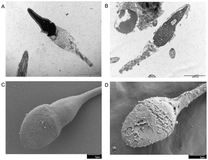
Figure 7. The microscopic ultrastructural changes in the sperm.
Transmission electron micrographs of human sperm samples incubated in the absence or presence of DGT (at MEC). (A) Control spermatozoa show proper acrosomal cap with intact plasma membrane, while (B) DGT-treated spermatozoa exhibit dissolution of the acrosomal cap. High resolution scanning electron micrographs (×15000 and ×19000) of human sperm treated without and with DGT at MEC. (C) Control sperm shows intact acrosomal cap and plasma membrane around the head and neck regions, while (D) DGT-treated sperm demonstrates dissolution of the acrosomal cap.

